Home » Without Label » Muscles In The Body Diagram - Muscles Chart Description Muscular Body Woman Muscle Chart With Accurate Description Of The Most Important Muscles Of The Canstock
Muscles In The Body Diagram - Muscles Chart Description Muscular Body Woman Muscle Chart With Accurate Description Of The Most Important Muscles Of The Canstock
Muscles In The Body Diagram - Muscles Chart Description Muscular Body Woman Muscle Chart With Accurate Description Of The Most Important Muscles Of The Canstock. Femur, capsule of knee, head of fibula. We hope this post inspired you and help you what you are looking for. To get started, choose a muscle group either on the muscle chart or in the muscle list on this page. It also helps raise the body from a supine. The muscular system is an organ system consisting of skeletal, smooth and cardiac muscles.
But, your soleus muscle in your lower leg and muscles in your back involved in maintaining posture contain mainly slow twitch muscle fibres. Labeled vector illustration chart on white background. Smooth muscle contractions are involuntary movements triggered by. Located immediately below the skin) muscles of the body. Use the location, shape and surrounding structures to help you.
Muscles Of The Trunk Anatomy Diagram Pictures Kenhub from thumbor.kenhub.com This is what happens in the body. These muscles hold the inner ear together and are connected to. There are some 700 named muscles in the body, and other smaller muscle tissues that are part of. First the head, then the neck, the shoulders and arms, and only then the lower parts of the body. Their main function is contractibility. The superficial back muscles are the muscles found just under the skin. Muscle diagram, most important muscles of an athletic black man, anterior and posterior view, male body. Found only in the heart, cardiac muscle is responsible for pumping blood throughout the body.
See how all sharpness disappears?
In the muscular system, muscle tissue is categorized into three distinct types: Almost every movement in the body is the outcome of muscle contraction. Located immediately below the skin) muscles of the body. Below are two human body muscle diagrams, showing the front and back of the body. But, your soleus muscle in your lower leg and muscles in your back involved in maintaining posture contain mainly slow twitch muscle fibres. Calcaneus tibial nerve plantar flexes foot. Their main function is contractibility. Muscle diagrams are a great way to get an overview of all of the muscles within a body region. Heaviest muscle in body, extends/straightens leg at hip during walking. The human muscular system is complex and has many functions in the body. Teres major is a thick and ovoid muscle in the upper arm. See how all sharpness disappears? This image is titled muscles of the body diagram picture and is attached to our article about 3 main muscle types in the human body.
These muscles hold the inner ear together and are connected to. Calcaneus tibial nerve plantar flexes foot. A muscle in the thigh that helps to rotate the leg into the sitting position assumed by a tailor. Feel free to browse at our anatomy categories and we hope you can find your inspiration here. See how all sharpness disappears?
Muscular System Anatomy And Physiology Nurseslabs from nurseslabs.com Published december 28, 2017 at 768 × 1024 in so…what can you feel or move? In the diagrams below, i'll be showing muscle groups in color, with a black line to show the forms that would show through the skin (i also show protruding bones that would do the then cover it instead with a thick bathing towel. Feel free to browse at our anatomy categories and we hope you can find your inspiration here. Labeled vector illustration chart on white background. It permits movement of the body, maintains posture and circulates blood throughout the body. This is a table of muscles of the human anatomy. The superficial back muscles are the muscles found just under the skin. See how all sharpness disappears?
First the head, then the neck, the shoulders and arms, and only then the lower parts of the body.
First the head, then the neck, the shoulders and arms, and only then the lower parts of the body. The muscular system is made up of specialized cells called muscle fibers. This is what happens in the body. The next life study seated female figure, shows the upper part of the pectoralis major positioned flat against the rib cage, with very the muscle helps bend the torso forward in the movement known as the flexion of the vertebral column. Found only in the heart, cardiac muscle is responsible for pumping blood throughout the body. The interactive muscle anatomy diagram shown below outlines the major superficial (i.e. The ear contains the smallest muscles in the body alongside the smallest bones. The muscular system is an organ system consisting of skeletal, smooth and cardiac muscles. There are approximately 640 skeletal muscles within the typical human, and almost every muscle constitutes one part of a pair of identical bilateral muscles, found on both sides, resulting in approximately 320 pairs of muscles, as presented in this article. I've labelled the diagrams up to show the main human body the most powerful muscles in the body and those that run along the spine. Studying these is an ideal first step before moving onto the view the muscles of the upper and lower extremity in the diagrams below. There are some 700 named muscles in the body, and other smaller muscle tissues that are part of. These muscles hold the inner ear together and are connected to.
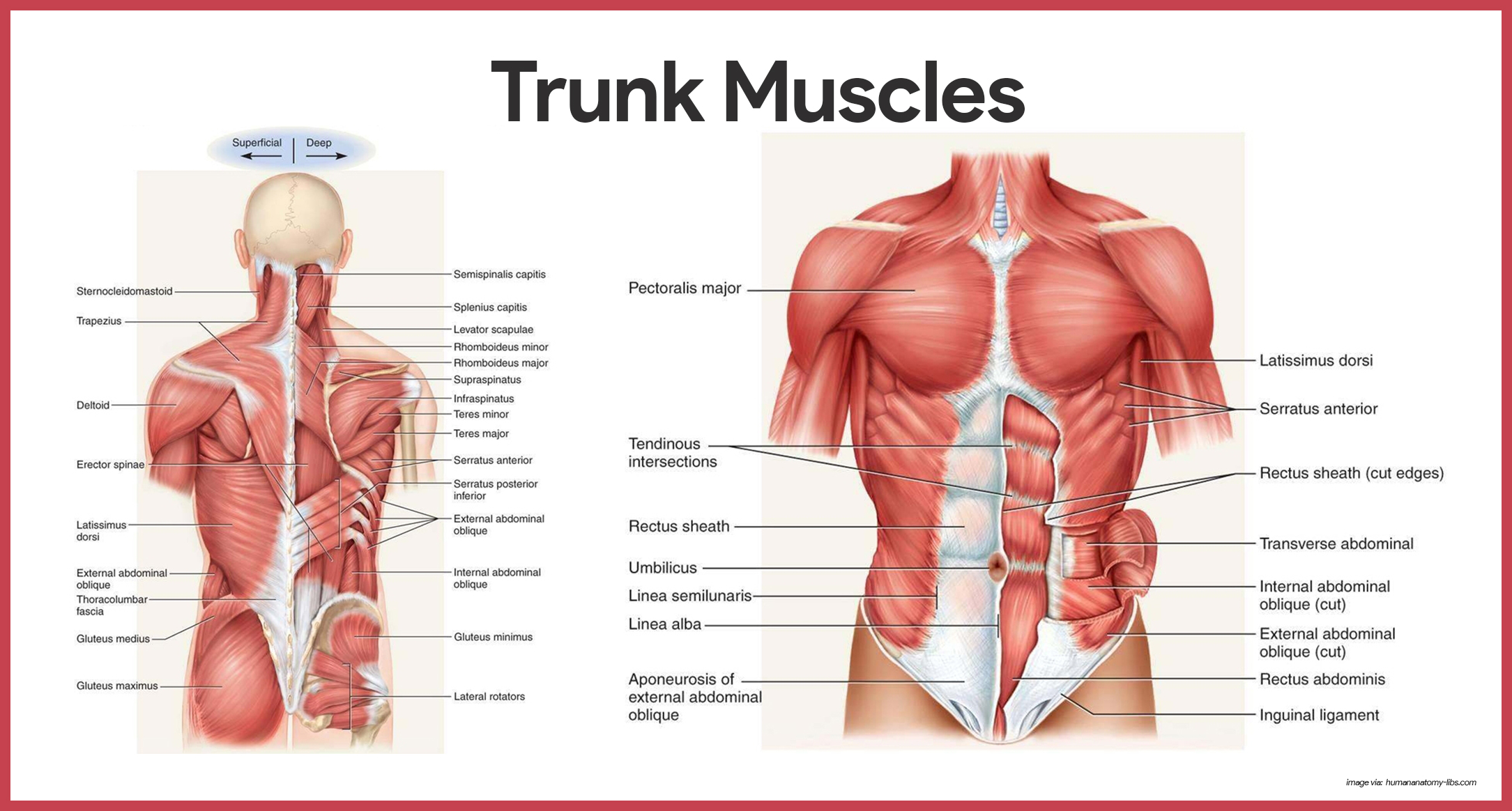
In this image, you will find frontalis, orbicularis oculi, zygomaticus, masseter, orbicularis oris, sternocleidomasteoid. Muscles, connected to bones or internal organs and blood vessels, are in charge for movement. This is a table of skeletal muscles of the human anatomy. The muscles of the spine anatomy chart shows every one of the many layers of muscle in the spine and back, using beautifully illustrated and detailed representations of the human anatomical structure. Thank you for visiting major muscles of the body diagram pictures.
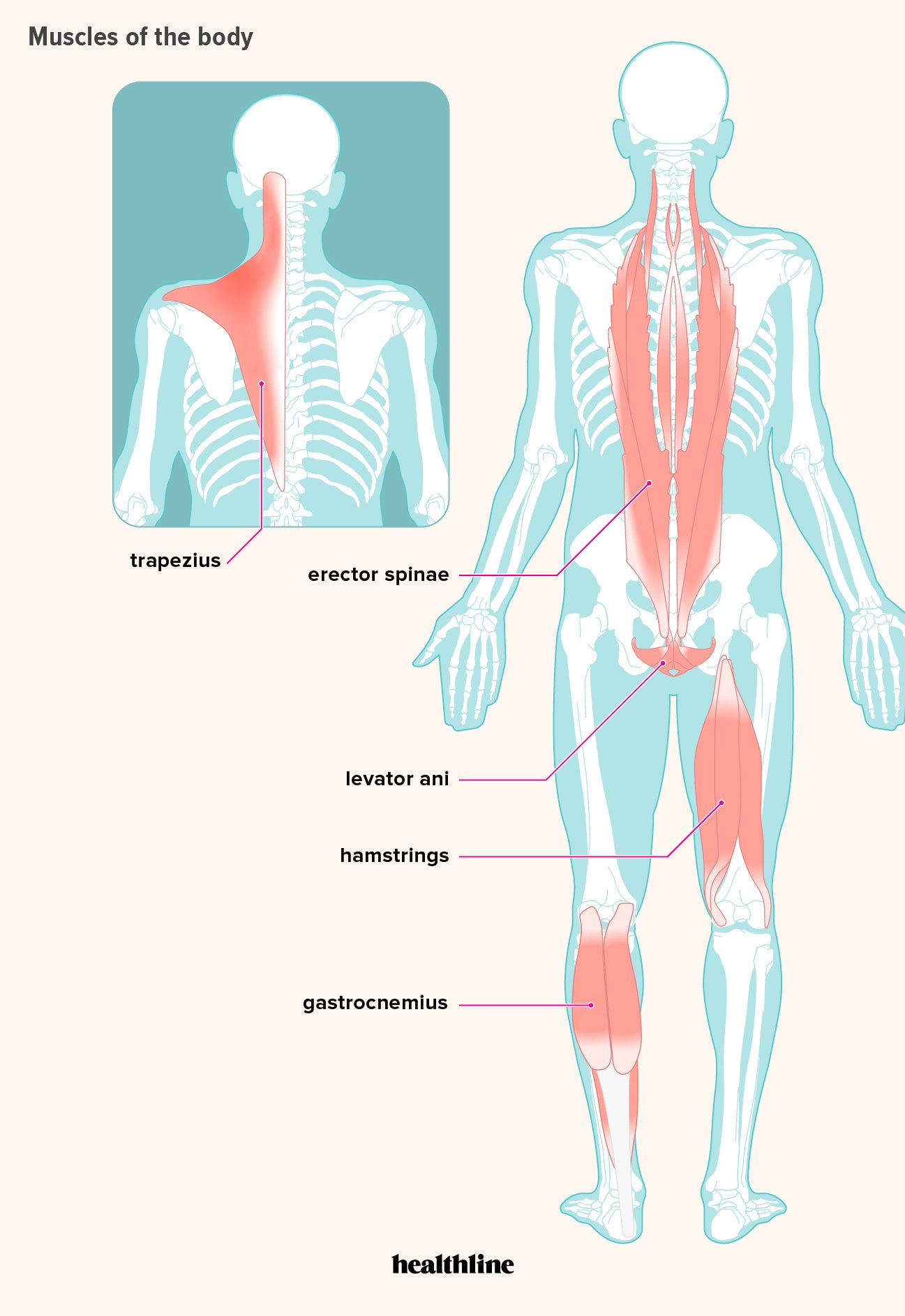
How Many Muscles Are In The Human Body Plus A Diagram from post.healthline.com Located immediately below the skin) muscles of the body. Their main function is contractibility. These muscles hold the inner ear together and are connected to. The ear contains the smallest muscles in the body alongside the smallest bones. Thank you for visiting major muscles of the body diagram pictures. Gas trocsoleus (gastrocnemius and soleus muscles). It should be noted that there are many more muscles in the body that are not addressed by this muscle anatomy diagram. There are some 700 named muscles in the body, and other smaller muscle tissues that are part of.
Anterior muscles in the body.
Diagrams of the muscles and guide to how they work. These muscles hold the inner ear together and are connected to. The sartorius muscle is positioned more superficially than the other in the leg muscles. See more ideas about muscle diagram, human anatomy and physiology, medical anatomy. This muscle diagram is interactive: The interactive muscle anatomy diagram shown below outlines the major superficial (i.e. Found only in the heart, cardiac muscle is responsible for pumping blood throughout the body. Labeled vector illustration chart on white background. See more ideas about body diagram, muscle anatomy, muscles in your body. Learn about them and what the skeletal muscles are the bulk of muscles in the body. This is a table of muscles of the human anatomy. Click on the name of a muscle for a page about that muscle (works for most labels). To get started, choose a muscle group either on the muscle chart or in the muscle list on this page.